
Digital Radiography

Clearer images with far less radiation

Digital radiography replaces traditional dental film with electronic sensors that capture high-resolution images of your teeth, roots, and supporting bone in seconds. The images appear instantly, can be enlarged and enhanced on screen, and are stored securely in your electronic record, no chemical processing, no waiting, and no repeat visits just to review results. Paired with intraoral cameras, digital X-rays let you see exactly what we see, making diagnoses easier to understand.

Sharper images, earlier diagnoses

Digital sensors capture fine detail that helps us find problems early, small cavities between teeth, failing restorations, root fractures, and bone changes associated with gum disease. Software tools let us adjust contrast, zoom in on areas of concern, and compare images side by side, so subtle problems are less likely to be missed.

Because images are easy to archive and retrieve, we can track changes over time. That historical view helps us tell the difference between stable findings and progressing conditions, informing decisions about whether to monitor or treat. Catching problems early usually means smaller, less invasive treatment.

Less radiation, more comfort

Digital sensors require substantially less radiation than traditional film while delivering equal or better image quality, especially important for children, pregnant patients, and anyone who needs frequent monitoring. Shorter exposure times and fewer retakes also make the experience faster and more comfortable.

We follow established, evidence-based guidelines for when and how often X-rays are appropriate, so every image taken directly supports a clinical decision or monitoring plan.

Better planning and communication

Because images appear instantly, we can review findings with you during the same appointment, explaining any concerns with the picture right in front of you. Digital imaging supports a wide range of care, including:

  • Routine checkups and cavity detection
  • Root canal evaluation and treatment planning
  • Implant planning, especially when combined with cone-beam CT and intraoral scans
  • Monitoring gum and bone health over time

When collaborative care is needed, high-quality digital files can be shared securely with dental labs and specialists, so everyone involved is working from the same clear picture.

A core part of modern, patient-centered care

Digital radiography integrates with intraoral cameras and electronic records to create one cohesive picture of your oral health. Enlarged, annotated images shown on a monitor make complex anatomy understandable, which helps you participate in treatment decisions rather than just receive them. Eliminating film and chemical processing also reduces waste and keeps appointments focused on your care instead of paperwork.

The cumulative result is clearer diagnostics, faster visits, lower radiation exposure, and more informed conversations about your options. If you’d like to learn more about how digital imaging fits into your dental care and what to expect at your next visit, please contact us.
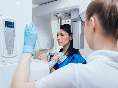
Digital radiography, Kary Dental

Our Office

Frequently Asked Questions

What is digital radiography?

Digital radiography is a modern method of capturing dental images using electronic sensors and computer software instead of traditional film. The sensor records the internal structures of the teeth and jaws and converts that information into a digital image that can be viewed on a monitor. These images provide high-resolution detail that helps dental professionals evaluate teeth, bone, and surrounding tissues.

Because images are produced and stored electronically, they can be manipulated for better clarity, enlarged for close inspection, and archived in the patient record without physical storage space. Digital radiography is used routinely for exams, restorative planning, and postoperative review as part of comprehensive dental care. Practitioners rely on these images to make informed diagnostic and treatment decisions efficiently.
Unlike traditional film x-rays, digital radiography captures images on a digital sensor rather than on photographic film that requires chemical development. Digital images appear instantly on a computer screen, eliminating the waiting period for film processing and reducing the chance of lost or damaged films. The digital workflow also allows immediate comparison of current and previous images for clearer clinical assessment.

Digital technology permits image enhancement such as contrast adjustment, magnification, and measurement tools that are not available with film. These capabilities improve visualization of fine details and support more accurate diagnoses. Additionally, digital images can be shared electronically with specialists or insurance carriers when clinically necessary, streamlining collaboration and referrals.
Digital radiography significantly reduces radiation exposure compared with conventional film x-rays because modern sensors are more sensitive and require lower doses of radiation to produce diagnostic-quality images. Safety protocols, such as lead aprons and thyroid collars when appropriate, remain standard practice to further minimize exposure. Clinicians follow guidelines that balance diagnostic necessity with the principle of using the lowest radiation dose reasonably achievable.

For most routine dental evaluations, the radiation dose from digital dental x-rays is very low and comparable to natural background radiation experienced over a short period. Pregnant patients should always inform their dental provider so appropriate precautions can be taken and imaging deferred when possible. The practice prioritizes patient safety and employs digital radiography to keep radiation exposure as low as possible while maintaining diagnostic effectiveness.
The two primary sensor types used in digital dentistry are direct digital sensors (solid-state detectors) and phosphor plate systems, often called PSPs. Direct digital sensors capture the image and transmit it immediately to a computer, providing near-instant results, while PSP plates are scanned after exposure to produce a digital file. Each system has clinical advantages related to image quality, patient comfort, and workflow preferences.

Direct sensors typically deliver faster results and higher immediate image clarity, which can speed chairside decision-making, while PSP systems are often thinner and more flexible for patient comfort. Clinicians select sensors based on the type of exam, patient anatomy, and the office’s workflow needs. Regular calibration and maintenance of sensors are essential to preserve image quality and reliable performance.
Digital x-rays enhance diagnostic accuracy by providing clear, high-resolution images that can be adjusted for brightness, contrast, and magnification to reveal subtle clinical details. These enhancements assist clinicians in detecting cavities, assessing bone levels, locating root anatomy, and identifying pathologies that may be invisible on a visual exam alone. Improved visualization supports more precise treatment planning across restorative, periodontal, endodontic, and surgical procedures.

Because digital images integrate easily with practice management and imaging software, they can be used alongside digital impressions, 3D scans, and intraoral camera photos to create comprehensive treatment plans. This integrated approach allows clinicians to simulate outcomes, evaluate alternative options, and communicate findings effectively with patients. At Kary Dental, these capabilities help the team present evidence-based recommendations and coordinate care with specialists when needed.
A digital radiography appointment is typically quick and minimally invasive, involving placement of a small sensor inside the mouth or positioning an external sensor for extraoral images. Patients may feel minor pressure from the sensor or bite tab, but the process is fast and designed for comfort. The clinician will position the sensor carefully and step behind a protective barrier or leave the room while the image is captured.

After exposure, the image appears on the computer screen within seconds and can be reviewed immediately with the patient to explain findings. This instant feedback improves communication and allows the dentist to answer questions and discuss treatment options during the same visit. The streamlined workflow reduces overall chair time while maintaining diagnostic thoroughness.
Digital x-rays are stored as part of the patient’s electronic health record on secure practice software that supports HIPAA-compliant data management and backups. Images are indexed to the patient file for easy retrieval during future visits and are retained according to professional record-keeping standards. Secure storage reduces the need for physical archives and simplifies long-term access to historical images for comparison.

When clinically necessary, digital images can be shared electronically with other dental offices or specialists using encrypted transfer methods or secure portals to protect patient privacy. Patients who request copies for continuity of care may be provided digital files or printed images as allowed by privacy regulations. The office of Kary Dental follows established protocols to safeguard imaging data while enabling appropriate clinical collaboration.
Yes, digital radiography is frequently used in conjunction with other imaging modalities such as cone-beam computed tomography (CBCT), intraoral cameras, and digital impressions to provide a more complete view of oral structures. Each technology contributes different types of information, CBCT provides three-dimensional bone detail, intraoral cameras show surface conditions, and digital impressions capture soft-tissue contours. Combining these tools enhances diagnostic precision and supports complex treatment planning.

Integration is facilitated by imaging software that allows side-by-side comparisons and overlays, enabling clinicians to correlate 2D and 3D data efficiently. This multimodal approach is particularly valuable for implant planning, endodontic assessment, and advanced restorative cases. Clinicians coordinate the appropriate imaging combination based on the clinical question and patient needs.